Immediate action: Stop manual handling and support the spinal region; apply cold packs 15–20 minutes every 2–3 hours for the first 48–72 hours, then switch to moist heat 15–20 minutes twice daily if pain and stiffness persist. For short-term analgesia consider ibuprofen 200–400 mg every 4–6 hours as needed (OTC limit 1,200 mg/day) or acetaminophen 500–1,000 mg every 4–6 hours (limit 3,000 mg/day); review contraindications and prescription history before combining medications.
Red flags requiring urgent assessment: progressive lower-extremity weakness, new perineal numbness, sudden loss of bladder or bowel control, fever above 38°C, or pain that fails to respond to appropriate medication. These findings warrant immediate clinical evaluation, neurological testing and possible imaging.
Early recovery plan: begin short walking intervals (10–20 minutes) multiple times per day within the first 48–72 hours as tolerated. Introduce low-load motor control work after 72–96 hours (pelvic tilts, transverse-abdominis activation, glute bridges). Reintroduce manual loads around the two-week mark starting with very light carries (under ~10% of body weight) using strict hip-hinge mechanics; increase load by ~10–20% per week only if pain-free and function improves.
Handling technique and prevention: keep objects close to the torso, maintain a neutral spine, initiate movement with hips and knees rather than the trunk, avoid twisting under load, and exhale while bracing the core. Use wheeled transport or team lifts for bulky items and schedule regular microbreaks when repetitive handling is unavoidable.
Reassessment criteria for progressive duties: consistent pain reduction at rest and during controlled practice lifts, near-equal strength bilaterally, normal gait pattern, and absence of new neurological signs. If recovery plateaus or neurological symptoms develop, arrange specialist referral and supervised rehabilitation focused on graded exposure and load tolerance testing.
Delayed lumbar strain after hauling suitcases: evidence-based guidance
Immediate actions: rest 24–48 hours; apply cold pack 15–20 minutes every 2–3 hours during the first 48–72 hours, then transition to moist heat and gentle mobility; analgesic options – ibuprofen 200–400 mg every 6–8 hours (maximum 1,200 mg/day OTC) or acetaminophen 500–1,000 mg every 4–6 hours (observe product and medical limits).
Warning signs requiring urgent assessment
- Progressive leg weakness or new difficulty walking
- Numbness/tingling in saddle distribution (inner thighs, perineum)
- New urinary retention or loss of bowel control
- High fever or worsening focal tenderness after significant trauma
Structured self-care and rehabilitation
- First 48–72 hours: relative rest with frequent short walks to maintain circulation; avoid prolonged bed rest.
- After 48–72 hours: begin gentle lumbar mobility (pelvic tilts, knee‑to‑chest, cat–cow), 10–15 minutes twice daily as tolerated.
- Weeks 1–3: progressive core activation (dead bug, bird‑dog), glute bridges and hip mobility drills; 2–3 sessions weekly, progressing by symptom response.
- Refer for physical therapy if functional limitation persists beyond 2 weeks or symptoms recur with routine tasks; typical program 6–8 weeks emphasizing motor control and graduated load tolerance.
- Medication adjuncts: topical NSAID preparations, short course muscle relaxant for severe spasm per prescriber; consider image‑guided steroid injection only for confirmed radicular irritation unresponsive to conservative care.
Practical prevention: split contents into multiple wheeled cases, use backpack with hip belt, position cases at waist height before raising, hinge at the hips with a neutral spine, avoid torso rotation while elevating bags, request assistance for bulky or awkward parcels.
Which specific injuries commonly cause delayed-onset spinal pain after carrying bulky suitcases
Recommendation: Obtain medical evaluation within 48–72 hours when progressive neurological deficits (leg weakness, numbness, saddle anesthesia) or bladder/bowel changes follow transporting weighty suitcases.
Herniated lumbar disc (disc prolapse): Nucleus pulposus extrusion compressing a nerve root produces radicular pain, paresthesia and possible motor deficit. Symptom onset ranges from immediate to delayed; classic radiculopathy often peaks within 24–72 hours. MRI is the diagnostic standard (sensitivity for symptomatic extrusion ~85–95%). Initial management: oral NSAIDs, short activity modification, targeted physiotherapy; refer for epidural steroid injection or surgical decompression when progressive motor loss or cauda equina signs appear.
Paraspinal muscle strain or tear: Localized posterior muscle overload from manual handling or prolonged carrying of bulky bags leads to focal tenderness, spasm and limited motion. Typical recovery spans 1–6 weeks with progressive improvement using analgesics, heat, phased stretching and strengthening guided by a physiotherapist. Ultrasound helps identify hematoma or sizable soft-tissue defects.
Facet joint injury and capsular irritation: Axial rotation combined with asymmetric load may sprain facet capsules, producing localized, activity-dependent pain that can mimic discogenic symptoms. Diagnostic intra-articular or medial branch block confirms the source; management includes mobilization, targeted exercise and steroid injection for persistent pain.
Sacroiliac joint dysfunction: One-sided carrying stresses the sacroiliac articulation, causing referred posterior pelvic and lower-extremity pain. Provocative tests (FABER, distraction) plus diagnostic SI injection establish diagnosis. Treatment: manual stabilization, pelvic core strengthening and brief external support for instability.
Vertebral compression fracture: Axial overload or sudden flexion in patients with low bone density may cause wedge fracture with focal pain that can worsen over time. Plain radiographs detect most fractures; MRI identifies marrow edema and acuity. Management options range from analgesia and spinal bracing to vertebral augmentation when pain is refractory or deformity progresses.
Nerve root contusion, epidural hematoma or spinal infection: Traction or direct insult to neural elements may produce delayed worsening of radicular symptoms; epidural hematoma often presents with rapid decline, while epidural abscess typically adds fever and systemic signs. Urgent MRI and labs (CBC, CRP, blood cultures) are indicated when systemic symptoms or rapidly evolving neurologic deficits appear; early surgical or infectious-disease input reduces risk of permanent impairment.
Red-flag indicators for urgent imaging/referral: new bilateral leg weakness, saddle numbness, urinary retention or incontinence, unexplained fever, or marked loss of mobility. Absent red flags, structured conservative care with analgesia, progressive mobilization and physiotherapy is appropriate; escalate evaluation if no meaningful improvement within one week or if neurologic signs develop.
Note: If vehicle upholstery was soiled during transport of packed cases, stain- and odor-removal guidance is available at how to clean cat pee from car seat.
How to tell a delayed muscle strain apart from nerve or disc-related pain
Treat a localized, tender lumbar muscle injury with conservative measures; seek prompt neurological assessment when radicular symptoms, progressive motor loss, or sphincter disturbance are present.
Key clinical distinctions
Muscle strain: pain usually felt as a diffuse ache or sharp focal tenderness in the lumbar muscles, worse with resisted contraction or trunk flexion/extension, reproducible with direct palpation, and commonly peaks 24–72 h after the precipitating event. Active range of motion is limited more than passive range. Pain that responds to ice initially and heat after 48–72 h, and improves with gradual loading and targeted stretching, aligns with a muscular origin. Expected recovery for uncomplicated strains is typically 2–6 weeks.
Radicular/disc-related pain: lancinating, electric, or burning pain that radiates below the knee or follows a dermatomal pattern suggests nerve-root involvement. Sensory loss, paresthesia, objective motor weakness, or reflex changes indicate neural compromise. Reproduction of symptoms with straight-leg raise at approximately 30–70° supports lumbar nerve root tension; coughing, sneezing or Valsalva-induced worsening implies intrathecal pressure increase from a disc lesion. Symptoms that persist beyond 6 weeks or worsen neurologically warrant advanced imaging and specialist input.
Objective signs, tests and thresholds for escalation
Sensory and motor mapping: L4 often affects medial shin and knee extension (quadriceps); L5 commonly involves dorsum of foot and great toe dorsiflexion; S1 typically produces lateral foot symptoms and reduced ankle plantarflexion with diminished Achilles reflex. A new motor deficit graded ≤3/5 requires urgent referral. A positive crossed straight-leg raise increases suspicion for significant disc herniation.
Red flags requiring immediate imaging or emergency evaluation: saddle anesthesia, acute urinary retention or fecal incontinence, rapidly progressive bilateral lower-limb weakness, fever with spine pain, history of malignancy or recent major trauma. For radicular pain without red flags, obtain MRI if severe functional impairment persists beyond ~6 weeks or if progressive neurological signs develop sooner.
Initial management differences: strains–short course of NSAID or acetaminophen for analgesia (ibuprofen 200–400 mg every 4–6 h as needed; naproxen 220 mg every 8–12 h), topical analgesics, early gentle mobilization and targeted physiotherapy; neural compression–consider early MRI, specialist-directed options such as epidural steroid injection for refractory radicular pain, and surgical decompression for progressive or severe deficits. Document focal findings, repeat neurological exam at follow-up, and escalate based on objective deterioration rather than pain intensity alone.
Practical at-home measures to reduce delayed lumbar pain after manual load
Begin cold therapy: apply an ice pack (wrapped in a thin cloth) to the lumbar region for 15–20 minutes every 2–3 hours during the first 48–72 hours to limit inflammation and swelling.
Transition to moist heat after the initial 48–72-hour period: use a warm compress or heating pad for 15–20 minutes before gentle mobility work to increase tissue extensibility and reduce stiffness.
Oral analgesia guidance: acetaminophen 500–1,000 mg every 4–6 hours (maximum ~3,000 mg/day); ibuprofen 200–400 mg every 4–6 hours (OTC max ~1,200 mg/day); naproxen 220 mg every 8–12 hours (OTC max ~660 mg/day). Avoid NSAIDs if on anticoagulants or with active peptic ulcer/kidney disease; consult a clinician for chronic liver disease before using acetaminophen.
Topical options: diclofenac 1% gel applied 3–4 times daily; 5% lidocaine patches applied up to 12 hours per day; menthol or capsaicin creams for localized relief. Do not apply heat over recently applied topical counterirritants that produce warmth.
Maintain gentle movement rather than prolonged immobilization. Aim for two 10–20-minute walks daily and perform spinal mobility drills: pelvic tilts 10–15 reps, cat–camel 8–10 reps, bird‑dog 8–12 reps per side, supine knee-to-chest 10 reps. Hold static hamstring stretches 30 seconds ×3 per leg. Progress loading gradually and avoid sudden twists or strenuous manual tasks for at least one week.
Posture and sleep adjustments: when seated, use a lumbar roll or small towel, keep hips slightly above knees, and rest feet flat. For sleep, side-lying with a pillow between knees or supine with a pillow under knees reduces lumbar strain. Prefer a medium-firm mattress and avoid slumped reclining positions for prolonged periods.
Self-massage and soft-tissue work: use a tennis ball against a wall for 1–2 minutes on tender spots, gentle foam-roller glute/hamstring work, or low-setting percussive devices for 1–2 minutes per muscle group. Stop if numbness or increased tingling develops.
Temporary external support: an elastic lumbar brace may permit safer activity for short-term symptomatic relief; limit continuous brace use to 48–72 hours to reduce risk of core deconditioning.
Escalation criteria: obtain urgent medical assessment with progressive lower-extremity weakness, new saddle-area numbness, loss of bowel or bladder control, fever above 38°C (100.4°F), or intractable pain unresponsive to rest and medication. Arrange outpatient clinician or physical therapy review if no meaningful improvement within one to two weeks or if radicular symptoms persist beyond one week.
Warning signs and timelines: when delayed spinal symptoms after handling bulky loads require medical attention
Seek urgent medical assessment for new unilateral or bilateral leg weakness, loss of bladder or bowel control, saddle numbness, or rapidly progressing sensory loss following onset.
Emergency red flags
Any of the following mandates immediate evaluation: new bowel or bladder dysfunction (retention or incontinence); saddle-region anesthesia; rapidly worsening motor deficit in one or both legs; inability to stand or walk unassisted; fever >38°C together with localized spinal pain; visible deformity after trauma suggesting fracture; sudden severe pain with known malignancy or prolonged corticosteroid use.
Timelines, thresholds and investigations
Neurological deficit present: obtain MRI within 24–72 hrs; treat as potential cauda equina or compressive myelopathy. Suspected fracture after notable trauma or high-risk bone disease: plain radiographs immediately, CT if radiographs inconclusive. Persistent severe pain without neurologic signs: seek primary care if no improvement within one week despite conservative measures; refer to spine specialist if symptoms persist beyond six weeks or if function remains markedly limited. Analgesic failure threshold: pain score ≥7/10 despite combined acetaminophen and NSAID, or inability to ambulate safely. Infection workup: order CBC, CRP, ESR and blood cultures when fever, night pain, elevated inflammatory markers, or recent systemic infection occur. Malignancy suspicion (history of cancer, unexplained weight loss, age >50): expedite imaging and oncology/orthopedics referral. If MRI contraindicated, use CT plus targeted neurologic exam. Any progressive neurologic decline at any point requires emergent re-evaluation and likely surgical consultation.
Limit single-item carry to ≤15% of body mass; prefer wheeled transport and two-handed lifts with a hip hinge.
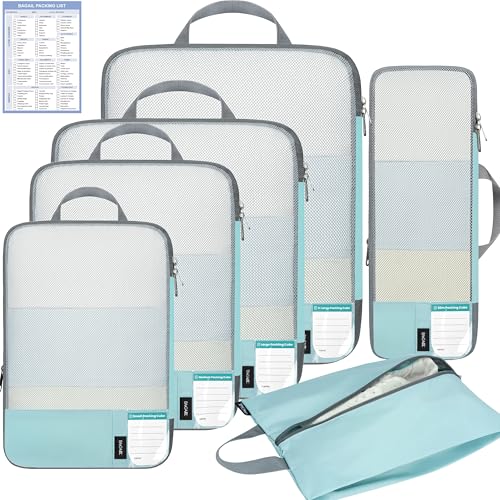
Pack distribution: place dense items closest to the suitcase’s or bag’s wheels or frame to keep the center of mass near the body’s midline. Use packing cubes to compress clothes and prevent weight shifting; position toiletries and shoes at the base. When consolidating for short trips, opt for multiple small bags rather than one bulky load. Compare selections and sizes at women’s toiletry picks and family-oriented economical options at budget family picks.
Handling and transfer technique
Squat with feet hip-width, hinge at hips and knees, and keep the load within 10–15 cm of the torso before rising. Avoid trunk rotation while lifting or setting down; pivot with the feet instead. When moving items between surfaces, perform two-person transfers for loads >20% body mass. Limit single static holds to under 15 seconds; rest the load on a surface when adjustments are required.
Travel movement, seating and conditioning
During transit, interrupt prolonged sitting every 45–60 minutes with 2–3 minutes of walking and light hip flexor/glute activation. In vehicles or aircraft, support the lumbar curve with a small cushion positioned at the beltline; tilt seat so hips sit slightly higher than knees. Pre- and mid-trip routines: plank series (3×30–60 s), bird-dogs (2×10 per side), and glute bridges (3×12) performed 2–3 times weekly improve load tolerance. Before any manual handling, warm up with 10 slow hip hinges and 10 cat–cow repetitions.
Equipment choices: choose four-wheel spinners for long terminal distances; select backpacks with padded hip belts and sternum straps if shoulders will bear load. For family travel, distribute contents across children’s or partner’s cases to reduce individual mass and peak force during lifts.
Quick preventive checklist: weight each piece at packing, redistribute dense items centrally, engage legs and hips when raising loads, walk every hour during long journeys, and maintain a short daily core routine starting two weeks before a major trip.
FAQ:
Can I develop back pain several days after I lifted heavy luggage?
Yes. Straining muscles, small tears in soft tissues, or irritation of spinal structures can cause pain that appears or worsens a day or two after the lifting event. Mild cases usually improve with short rest, cold packs for the first 48 hours, then heat, gentle walking, and over-the-counter pain relievers such as ibuprofen or acetaminophen. See a clinician promptly if you have severe pain that does not improve, numbness or weakness in a leg, loss of bladder or bowel control, fever, or pain that spreads down the leg.
What should I do if my back starts hurting days later and I need to travel again or lift bags?
If you must travel or handle luggage while your back is painful, take steps to reduce strain. Use wheeled bags and ask for help lifting or carrying heavy items. Keep loads low and close to the body, bend at the knees and hips instead of rounding the lower back, and avoid twisting while lifting. Continue short walks and light movement to keep muscles limber, and apply ice for acute inflammation or heat for tightness after 48 hours. Over-the-counter anti-inflammatory medication can reduce pain and swelling for many people; follow dosing directions and check with a clinician if you have other health conditions or take other drugs. If pain persists beyond two weeks, worsens, or is accompanied by numbness, weakness, or changes in bowel or bladder control, arrange a medical evaluation. A clinician may recommend targeted tests, a short course of supervised exercise or manual therapy, or referral to a spine specialist or physical therapist if recovery stalls.


